
Our Comprehensive Services
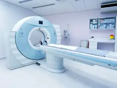
CT Scan
The 96 slice Multi detector CT scan Machine (MDCT), at Health Wave diagnostics, provides coverage, speed, power and resolution, which enables physicians to capture images of the body in few seconds, something which no other CT system can offer.
Special Features:
- High-Resolution Fast Imaging Of Any Body Region With Exquisite Multiplanar Reconstructions.
- Excellent 3-D Reconstruction Of Bones And Other Structures Showing Detailed Anatomy, Free Of Artefacts.
- Non-Invasive Evaluation Of Blood Vessels (Angiographies)
- Whole Body CT In Few Minutes With Sub-Millimeter Resolution.
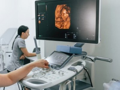
Hi-End Sonography
Ultrasound imaging, also called ultrasound scanning or sonography, involves the used of a small transducer (probe) and ultrasound gel to expose the body to hide- frequency sound waves. Ultrasound is a safe and painless, and produces pictures of the inside of the body using sound waves.
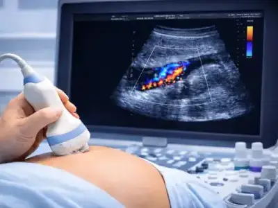
3D-4D Color Doppler
Ultrasound imaging, also called ultrasound scanning or sonography, involves the used of a small transducer (probe) and ultrasound gel to expose the body to hide- frequency sound waves.
Ultrasound Examinations Do Not Use Ionizing Radiation (As Used In X-Rays). Because Ultrasound Images Are Captured In The Real- Time, They Can Show The Structure And The Movement Of The Body’s Internal Organs, As Well As Blood Flowing Through Blood Vessels. Ultrasound Imaging Is A Noninvensive Medical Test That Helps Physician Diagnose And Treats Medical Conditions.
Conventional Ultrasound Displays The Images In Thin, Flat Sections Of The Body. Advancement In Ultrasound Technology Include Three- Diamensional (3-D) Ultrasound That Formats The Sound Wave Data Into 3-D Images. Four- Diamensional (4-D) Ultrasound Is 3-D Ultrasound In Motion. Color Doppler Ultrasound Is A Special Ultrasound Technic That Evaluates Blood Flow Through A Blood Vessel, Including The Body’s Major Arteries And Veins In The Abdomen, Arms, Legs And Neck.
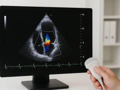
2D Echo Color Doppler
Diagnose a variety of heart conditions and assess damage after a heart attack or diagnose for valvular heart disease (2D Echo).
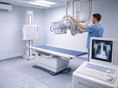
Digital X-Ray
Equipped with the latest digital mechines, Health wave Diagnostics ensures the best quality of X-rays & procedures. It includes routine radiography, special radiography, dental radiography – digital OPG, lateral cephalometry and occlusal view, IOPA also includes portable Digital X-Ray.
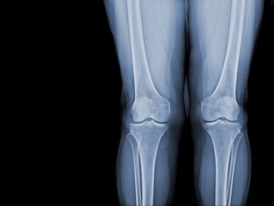
Full Length Scanogram
A method of making radiographs by the use of a narrow slit beneath the tube in such a manner that only a line or sheet of x-rays is employed and the x-ray tube moves over the object so that all the rays of the central beam pass through the part being radiographed at the same angle.
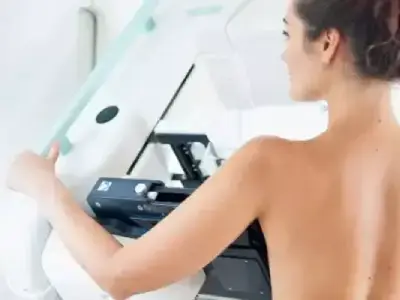
Digital Mammography
Mammography is important because in its earliest stages breast cancer may not be palpable it may be too small to feel as a lump or tissue change. Mammography can help detect these tissue changes two year or more before you would feel them.
Physical examination is also important because premenopausal breast tissue is often dense and fibrous, which may decrease the reliability of mammography for young women. If you notice any change in your breast, see your doctor. A biopsy may be essential for final diagnosis if a lump is detected.
Patient Benefits:
High resolution films so that even the smallest of cancer can be easily detected with least discomfort and automatic decompression.
Age Recommendations:
- Over 50 Years: Annual mammography examination.
- 40-50 Years: Every 1-2 years per physician guidance.
- Under 40 Years: Baseline mammogram may be done at 35 years.
Cardiac Imaging: Computerized Stress Test (TMT), 2D echo color doppler, ECG, Cardiology studies etc.
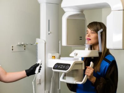
Dental CT Scan
Cone beam computed tomography (CBCT) generates three-dimensional (3D) images of dental structures, soft tissues, nerve paths and bone in the head and face region in a single scan for precise treatment planning.
Typical uses of CBCT:
- Dental implants: Location of anatomic structures, bone quality/quantity, implant placement.
- Oral & maxillofacial surgery: Impacted teeth, facial fractures, orthognathic planning.
- Pathology & Orthodontics: Lesions, cleft palate, root angulation.
- TMJ: Osseous structures and joint relationships.
Advantages: Lower radiation dose, open environment, 20-second scan, instant screen availability.
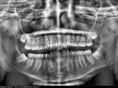
OPG & TMJ Imaging
CBCT reformatted panoramic images outperform digital panoramic images acquired through conventional OPG machines, providing an undistorted and accurate dimensional view of the jaws free of magnification and superimposition.
TMJ Imaging: Effective diagnostic assessment for temporomandibular joint conditions, detecting subtle osteoarthritic changes like cysts, sclerosis, and bone remodeling.
Lateral Cephalogram: Profile x-ray used in Orthodontics to assess tooth and jaw relationships.
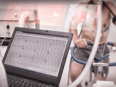
Computerized Stress Test & ECG
Computerized Stress Test: For stress test (TMT), 2D echo color doppler, ECG, cardiology studies, etc.
ECG Test: Simple test used to check your heart's rhythm and electrical activity via skin sensors detecting electrical signals produced during each heartbeat.
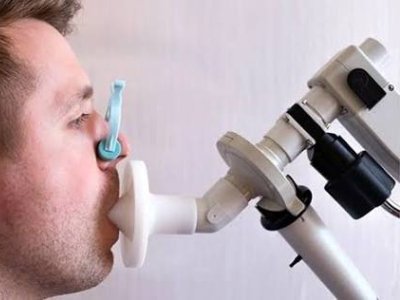
Pulmonary Function Test
Measures how well the lungs take in and release air and transfer gases like oxygen into circulation.
How it's performed: Spirometry measures airflow rate and volume. Lung volumes can also be measured via body plethysmograph or tracer gas dilution methods.
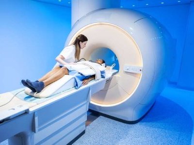
Whole Body High Definition MRI
Magnetic Resonance Imaging (MRI) visualizes internal body structures with excellent soft-tissue contrast without X-ray radiation, making it safe during pregnancy.
Covers brain, spine, joints, nerves, whole-body screening, MR cholangiopancreatography, MR urography, MR Defaecography, MR Enterography, and vascular angiographies.
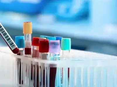
Advanced Pathology & Laboratory Services
Serology & Immunology: CLIA, ELISA, and rapid assays for HIV, Hepatitis, Typhoid, Dengue, Chikungunya, and hormone/tumor markers.
Clinical Pathology & Cytopathology: Routine analysis of body fluids, urine, stool, and FNA cytology.
Biochemistry & Haematology: Fully automated analysis for cardiovascular, renal, liver disorders, anaemias, and leukemias.
Allergy & Microbiology: Skin prick testing, IgE antibody screens, and automated Bact/ALERT & VITEK processing.
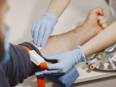
Specialized Diagnostic Packages
Integrated diagnostic care units aligned to support specialist physician consultations, preventative health screenings, senior citizen wellness, and cardiac evaluations.
Key Focus: Ensuring rapid turnaround times, high-precision reporting, and seamless integration between imaging and laboratory departments.